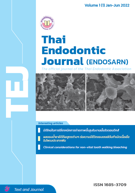
Effect of different EDTA formulae on dental pulp stem cells viability
Keywords:
รูปร่างเซลล์, ความมีชีวิตของเซลล์, เซลล์ต้นกำเนิดเนื้อเยื่อในโพรงประสาทฟัน, อีดีทีเอ, , สารลดแรงตึงผิวAbstract
Abstract
Objective: To study the effects of different EDTAs on cell viability of dental pulp stem cells (DPSCs) on root dentine.
Materials and method: Fifty-nine human extracted first mandibular premolar roots were sectioned bucco-lingually. Each half of root was cut vertically into a piece of dentine sample size 3.5X3.5X1 mm, which included root canal wall on one side. A total of 118 samples were randomly divided into 7 experimental groups of 16 each for submerging in one of the solutions for 1 minute: Endo Clean, Smearclear, Ultradent, 17% EDTA, 5.25% sodium hypochlorite, 0.9% Saline solution and DMEM culture medium as the Positive control. The negative control of 6 samples were in DMEM without cells. Three dentine samples from each experimental group were selected and examined the dentine surface with SEM. The remaining dentine samples were seeded with DPSCs and cultured. After 7 days, another 3 samples from each group were examined for cell morphology with SEM. The cell viability was determined by MTT assay from the remaining 10 samples.
Results: Percentage of cell viability in the groups of 17% EDTA, Ultradent, saline solution, and positive control were significantly higher than the Endoclean, Smearclear and 5.25% sodium hypochlorite groups (p<0.05). There was no significant difference between the groups of 17% EDTA, Ultradent, 0.9% saline solution and positive control and the Endoclean, Smearclear and 5.25% sodium hypochlorite groups (p>0.05). For dentine surfaces with SEM, the Endoclean group appeared to be mostly clear and large dentinal tubules, whereas the Smearclear group exhibited large dentinal tubules and some smear plugs. The Ultradent group presented tiny opened dentinal tubules with some smear plugs, while the EDTA group had medium opened dentinal tubules and some smear plugs. Heavy smear layer covering was observed in the sodium hypochlorite and the saline groups. For the 7 days of DPSCs culturing, the Endoclean group appeared round-shaped cells and a small number of spindle cells, whereas the Smearclear group exhibited only round cells. The Ultradent group presented mostly flat cells and small number of round and spindle cells, while the EDTA group had flat and round cells. The irregular shape cells and cell fragments were found only in the Sodium hypochlorite group. For the positive control group, the majority of cells were flat, the round- and spindle-shaped cells were rarely found.
Conclusion: In the condition of this study, cell viability of 17% EDTA Ultradent, 0.9% saline solution and positive control group was significantly higher than Endoclear, SmearClear and 5.25% sodium hypochlorite groups.
References
Murray PE, Garcia-Godoy F, Hargreaves KM. Regenerative endodontics: a review of current status and a call for action. J Endod. 2007; 33(4): 377-90.
Wigler R, Kaufman AY, Lin S, Steinbock N, Hazan-Molina H, Torneck CD. Revascularization: a treatment for permanent teeth with necrotic pulp and incomplete root development. J Endod. 2013; 39(3): 319-26.
Murray PE, Garcia-Godoy F, Hargreaves KM. Regenerative endodontics: a review of current status and a call for action. Journal of endodontics. 2007;33(4):377-90.
Wigler R, Kaufman AY, Lin S, Steinbock N, Hazan-Molina H, Torneck CD. Revascularization: a treatment for permanent teeth with necrotic pulp and incomplete root development. Journal of endodontics. 2013;39(3):319-26.
Rafter M. Apexification: a review. Dental traumatology : official publication of International Association for Dental Traumatology. 2005;21(1):1-8.
Garcia-Godoy F, Murray PE. Recommendations for using regenerative endodontic procedures in permanent immature traumatized teeth. Dental traumatology : official publication of International Association for Dental Traumatology. 2012;28(1):33-41.
Jeeruphan T, Jantarat J, Yanpiset K, Suwannapan L, Khewsawai P, Hargreaves KM. Mahidol study 1: comparison of radiographic and survival outcomes of immature teeth treated with either regenerative endodontic or apexification methods: a retrospective study. Journal of endodontics. 2012;38(10):1330-6.
Lovelace TW, Henry MA, Hargreaves KM, Diogenes A. Evaluation of the delivery of mesenchymal stem cells into the root canal space of necrotic immature teeth after clinical regenerative endodontic procedure. Journal of endodontics. 2011;37(2):133-8.
Gronthos S, Mankani M, Brahim J, Robey PG, Shi S. Postnatal human dental pulp stem cells (DPSCs) in vitro and in vivo. Proceedings of the National Academy of Sciences of the United States of America. 2000;97(25):13625-30.
Huang GT, Sonoyama W, Liu Y, Liu H, Wang S, Shi S. The hidden treasure in apical papilla: the potential role in pulp/dentin regeneration and bioroot engineering. Journal of endodontics. 2008;34(6):645-51.
Sonoyama W, Liu Y, Yamaza T, Tuan RS, Wang S, Shi S, et al. Characterization of the apical papilla and its residing stem cells from human immature permanent teeth: a pilot study. Journal of endodontics. 2008;34(2):166-71.
Huang X, Zhang J, Huang C, Wang Y, Pei D. Effect of intracanal dentine wettability on human dental pulp cell attachment. International endodontic journal. 2012;45(4):346-53.
Zhang R, Cooper PR, Smith G, Nor JE, Smith AJ. Angiogenic activity of dentin matrix components. Journal of endodontics. 2011;37(1):26-30.
Bettina Basrani, Haapasalo M. Update on endodontic irrigating solutions. Endodontic Topics. 2012;27:74-102.
Ring KC, Murray PE, Namerow KN, Kuttler S, Garcia-Godoy F. The comparison of the effect of endodontic irrigation on cell adherence to root canal dentin. Journal of endodontics. 2008;34(12):1474-9.
Galler KM, D'Souza RN, Federlin M, Cavender AC, Hartgerink JD, Hecker S, et al. Dentin conditioning codetermines cell fate in regenerative endodontics. Journal of endodontics. 2011;37(11):1536-41.
Yamada RS, Armas A, Goldman M, Lin PS. A scanning electron microscopic comparison of a high volume final flush with several irrigating solutions: Part 3. Journal of endodontics. 1983;9(4):137-42.
Rosenberg K, Olsson H, Morgelin M, Heinegard D. Cartilage oligomeric matrix protein shows high affinity zinc-dependent interaction with triple helical collagen. The Journal of biological chemistry. 1998;273(32):20397-403.
Roberts-Clark DJ, Smith AJ. Angiogenic growth factors in human dentine matrix. Archives of oral biology. 2000;45(11):1013-6.
Toledano M, Aguilera FS, Yamauti M, Ruiz-Requena ME, Osorio R. In vitro load-induced dentin collagen-stabilization against MMPs degradation. Journal of the mechanical behavior of biomedical materials. 2013;27:10-8.
Li R, Guo W, Yang B, Guo L, Sheng L, Chen G, et al. Human treated dentin matrix as a natural scaffold for complete human dentin tissue regeneration. Biomaterials. 2011;32(20):4525-38.
เลิศอักษรไพบูล วรงล. Effect of solution enchancing dental pulp stem cells on dentin attachment and cell survival. รายงานการวิจัยเพื่อประกาศนียบัตรชั้นสูง สาขาวิชาทันตแพทยศาสตร์ วิชาเอก วิทยาเอ็นโดดอนต์. 2012.
Pang N-S, Lee SJ, Kim E, Shin DM, Cho SW, Park W, et al. Effect of EDTA on Attachment and Differentiation
of Dental Pulp Stem Cells. Journal of endodontics. 2013:1-7.
AAE. Clinical Considerations for a Regenerative Procedure. 2013.
Hulsmann M, Heckendorff M, Lennon A. Chelating agents in root canal treatment: mode of action and indications for their use. International endodontic journal. 2003;36(12):810-30.
Song J, Liu L, Li P, Xiong G. Short-range interactions between surfactants, silica species and EDTA(4)- salt during self-assembly of siliceous mesoporous molecular sieve: a UV Raman study. Spectrochimica acta Part A, Molecular and biomolecular spectroscopy. 2012;97:616-24.
Bhatnagar R, M. DKN, Shivanna V. Decalcifying effect of three chilating agents. endodontology. 2006;18(2).
De-Deus G, Reis C, Fidel S, Fidel R, Paciornik S. Dentine demineralization when subjected to EDTA with or without various wetting agents: a co-site digital optical microscopy study. International endodontic journal. 2008;41(4):279-87.
Lui JN, Kuah HG, Chen NN. Effect of EDTA with and without surfactants or ultrasonics on removal of smear layer. Journal of endodontics. 2007;33(4):472-5.
Awad S, Allison SP, Lobo DN. The history of 0.9% saline. Clinical nutrition. 2008;27(2):179-88.
Barbosa SV, Barroso CM, Ruiz PA. Cytotoxicity of endodontic irrigants containing calcium hydroxide and sodium lauryl sulphate on fibroblasts derived from mouse L929 cell line. Brazilian dental journal. 2009;20(2):118-21.
Babich H, Babich JP. Sodium lauryl sulfate and triclosan: in vitro cytotoxicity studies with gingival cells. Toxicology letters. 1997;91(3):189-96.
http://en.wikipedia.org/wiki/Cetrimonium_bromide.
Spano JC, Barbin EL, Santos TC, Guimaraes LF, Pecora JD. Solvent action of sodium hypochlorite on bovine pulp and physico-chemical properties of resulting liquid. Brazilian dental journal. 2001;12(3):154-7.
Rajaraman R, Rounds DE, Yen SP, Rembaum A. A scanning electron microscope study of cell adhesion and spreading in vitro. Experimental cell research. 1974;88(2):327-39.
Al-Nazhan S. SEM observations of the attachment of human periodontal ligament fibroblasts to non-demineralized dentin surface in vitro. Oral surgery, oral medicine, oral pathology, oral radiology, and endodontics. 2004;97(3):393-7.
Downloads
Published
How to Cite
Issue
Section
License
Copyright (c) 2022 Thai Endodontic Journal

This work is licensed under a Creative Commons Attribution-NonCommercial-NoDerivatives 4.0 International License.
Thai Endod Journal is licensed under a Creative Commons Attribution-NonCommercial-NoDerivatives 4.0 International (CC BY-NC-ND 4.0) license, unless otherwise stated. Please read our Policies in Copyright for more information.


