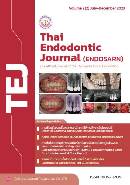
การทำศัลยกรรมปลายรากฟันร่วมกับการจัดการ ปุ่มกระดูกส่วนงอกของขากรรไกรที่มีขนาดใหญ่: รายงานผู้ป่วย
Keywords:
apicoectomy, endodontic surgery, exostosis, periosteal releasing incision, surgical endodontic retreatmentAbstract
Endodontic microsurgery is one approach for managing root canal treatment failures, especially when obstacles such as post and core with crown restorations are involved. However, cases with anatomical limitations, such as an exostosis impeding the surgical site, may result in more complex planning and treatment processes than usual. This case report aims to document a successful treatment procedure with an interdisciplinary approach to carry out a one-visit endodontic microsurgery concurrent with an exostosis removal in a 48-year-old Thai female patient. The patient presented with an acute apical abscess on her upper right central incisor due to a failure of the preceding primary root canal treatment. The tooth was previously restored with a post and core with crown, which remained in good condition with an intact margin. At the surgical site, a large exostosis was found to obstruct the endodontic surgical intervention. Moreover, the patient also had food impaction and cleaning difficulty in this area. As a result, the patient underwent apical surgery with the exostosis removal simultaneously. A one-year follow-up revealed that the patient had remained asymptomatic. The crown was still in good condition without any gingival recession. Furthermore, the size of the exostosis decreased, resulting in the patient’s improvement of oral hygiene care. Radiographic assessments confirmed a complete resolution of the periapical lesion.
References
Siqueira JF, Jr. Aetiology of root canal treatment failure: why well-treated teeth can fail. Int Endod J. 2001;34(1):1-10.
Friedman S, Stabholz A. Endodontic retreatment--case selection and technique. Part 1: Criteria for case selection. J Endod. 1986;12(1):28-33.
Bradford R.Johnson MIF, and Louis H.Berman. Periradicular surgery. In: Hargreaves KM, Berman LH, Rotstein I, Cohen S, editors. Cohen's pathways of the pulp. Twelth edition. St. Louis, Missouri: Elsevier; 2021. p. 433.
Smitha K, Smitha GP. Alveolar exostosis – revisited: a narrative review of the literature. Saudi J Dent Res. 2015;6(1):67-72.
Matthews DC, Sutherland S, Basrani B. Emergency management of acute apical abscesses in the permanent dentition: a systematic review of the literature. J Can Dent Assoc. 2003;69(10):660.
Kim S, Kratchman S. Modern endodontic surgery concepts and practice: a review. J Endod. 2006;32(7):601-23.
Tsesis I, Faivishevsky V, Kfir A, Rosen E. Outcome of surgical endodontic treatment performed by a modern technique: a meta-analysis of literature. J Endod. 2009;35(11):1505-11.
Urolagin S, Kale T, Patil S. Intraoral Incisions, design of flaps and management of soft tissue. Guident. 2010:57-61.
Stanley F Malamed D. Clinical action of specific agents. In: Stanley F Malamed D, editor. Handbook of Local Anesthesia. seventh edition. South Asia: Elsevier Health Sciences; 2019. p. 59, 213-4
Shillingburg HT HS, Whitsett LD, Jacobi R, Brackett SE. Preparations for extensively damaged teeth. In: Shillingburg HT, editor. Fundamentals of fixed prosthodontics. 3rd ed ed. Chicago: Quintessence Pub. Co. Chicago; 1997. p. 191-2.
Yildirim T, Er K, Taşdemir T, Tahan E, Buruk K, Serper A. Effect of smear layer and root-end cavity thickness on apical sealing ability of MTA as a root-end filling material: a bacterial leakage study. Oral Surg Oral Med Oral Pathol Oral Radiol Endod. 2010;109(1):e67-e72.
Chong BS, Ford TRP. Root‐end filling materials: rationale and tissue response. Endod Topics. 2005;11:114-30.
Torabinejad M, Pitt Ford TR. Root end filling materials: a review. Dent Traumatol. 1996;12(4):161-78.
Romanos GE. Periosteal releasing incision for successful coverage of augmented sites. a technical note. J Oral Implantol. 2010;36(1):25-30.
Griffin TJ, Hur Y, Bu J. Basic suture techniques for oral mucosa. Clin adv periodontics. 2011;1(3):221-32.
Ramachandran Nair PN, Pajarola G, Schroeder HE. Types and incidence of human periapical lesions obtained with extracted teeth. Oral Surg Oral Med Oral Pathol Oral Radiol Endod. 1996;81(1):93-102.
Schulz M, von Arx T, Altermatt HJ, Bosshardt D. Histology of periapical lesions obtained during apical surgery. J Endod. 2009;35(5):634-42.
Downloads
Published
How to Cite
Issue
Section
License
Copyright (c) 2023 Thai Endodontic Journal

This work is licensed under a Creative Commons Attribution-NonCommercial-NoDerivatives 4.0 International License.
Thai Endod Journal is licensed under a Creative Commons Attribution-NonCommercial-NoDerivatives 4.0 International (CC BY-NC-ND 4.0) license, unless otherwise stated. Please read our Policies in Copyright for more information.


